Situation
Company OR-CZ supports clients’ success by providing comprehensive services and cutting-edge software solutions, with focus on medical imaging and management and secure access to image data via a standard web browser from anywhere in the medical facility or the Internet.

Breast cancer is the most diagnosed type of cancer in women, and the leading cause of their death. Breast calcifications are small calcium deposits within breast tissue. Calcifications can be seen as white bright spots on mammograms. Even though most calcifications are benign, some patterns of calcifications groups or calcification shapes can signal malignancy (cancer). Each mammogram examination is marked with BIRADS category ranging from zero to six. If the calcification shape or pattern looks suspicious, the patient will be called for additional examination. These examinations are time exhausting, prone to human error and can vary in decisioning.
Solution
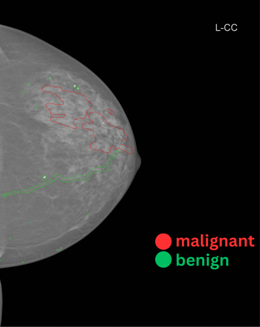
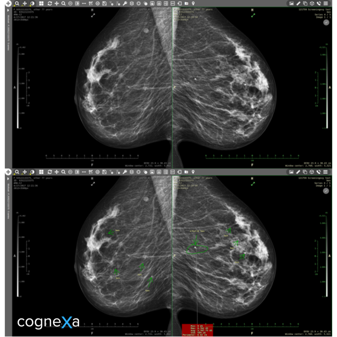
To address these issues, we developed an algorithm to automatically detect and classify calcifications in mammography studies. Results of this algorithm are pixel-wise detections – semantic segmentation technique originally proposed for medical imaging segmentation. Dataset was created in cooperation with Cognexa and OR-CZ. Consists of a set of mammograms with different BIRADS categories ranging from one to six. Mammograms were annotated and cross reviewed by experienced and attested mammologists. After that Cognexa and OR-CZ data scientists reviewed the quality of the annotation in the respect of machine learning applications. Multiple training experiments took place, varying in architectures, hyperparameters and dataset combinations before reaching the final model. Algorithm was designed in a way to help diagnosticians in searching for suspicious calcifications, occurrences and calcinations regions that could be under different circumstances overlooked, or/and create a second medical opinion.
Result
Our team created an algorithm with a U-Net backbone wrapped in docker images accessible via REST interface. Model was able to fulfill expectations and even find microcalcifications in a dataset that was supposed to be clear of calcifications. Module was hidden behind the FastAPI framework and containerized using Docker, for fast and stable deployment. The project also included integration into the MARIE PACS system, which forms a user interface for the use of artificial intelligence by doctors.


OR-CZ
Cognexa developed an algorithm to automatically detect and classify calcifications in mammography studies.
Tags
- Life Sciences
- MedTech
- Oncology
- Radiology
- Visual AI
- Workflow Automation


